Moderate weight bearing and minimal weight bearing exercise induce acute impact on collagen biochemical markers related to osteoarthritis
J.J. Bjerre-Bastos Osteoarthritis and Cartilage VOLUME 28, SUPPLEMENT 1, S63, APRIL 01, 2020
Purpose: Patients with osteoarthritis (OA) are recommended regular physical activity (PA) to limit pain and to preserve joint function, but the acute impact of joint loading on cartilage integrity in response to different types of PA (weight bearing vs non-weight bearing) remains to be explored. Joint-specific biochemical markers (BM) originating from type I-III collagen can be measured in serum and may reflect the turnover status of joint tissue. The aim of this study was to investigate the effect of running PA vs cycling PA on extracellular matrix (ECM) biomarkers reflecting collagens type I-III turnover. To our knowledge, this is the first study investigating the acute effect of exercise on the metabolism of joint related collagens in OA.
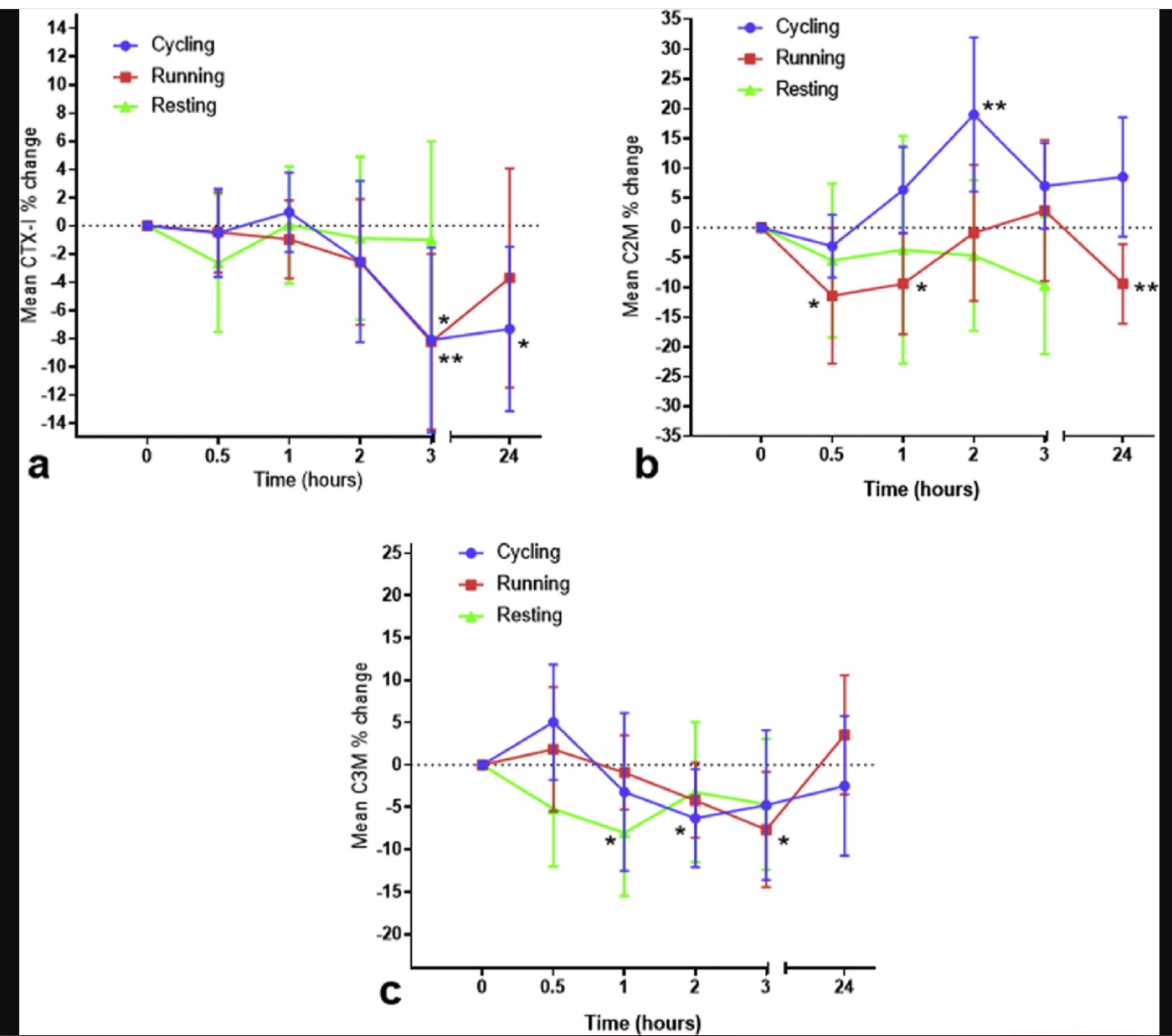
Methods: We conducted a randomized, cross-over clinical study including subjects with primary knee OA as determined by radiographic evaluation. Screening included a maximal heart rate (HRmax) test to establish physical capacity and to standardize exercise sessions at an intensity of approximately 75 % of the HRmax. Participants underwent running PA and cycling PA in randomized order with venous blood samples taken at baseline and 0.5, 1, 2 and 3 hours after exercise initiation and again 24 hours after the exercise in order to evaluate the dynamic levels of biomarkers. Potential diurnal variation was taken into account by measurements at comparable times from participants on a separate day with no exercise (resting). Levels of serum CTX-I (reflecting type I collagen turnover), C2M (reflecting type II collagen turnover) and C3M (reflecting type III collagen turnover) were measured by sandwich ELISA. The dynamics of biomarkers were plotted over time. Error bars represent 95% confidence intervals. Significance level was set to 0.05.
Results: 20 subjects were included of which 20 patients completed cycling and resting and 15 patients completed running. Figure 1a-c displays proportional biomarker changes from baseline. CTX-I decreased significantly from baseline at three hours after both running (p<0.01) and cycling (p<0.05) and was still decreased the day after running (p<0.05). No change in CTX-I levels was seen during rest. This suggests that exercise acutely reduces bone-turnover. C2M decreased significantly at 0.5 hour after running (p<0.05), but was found to be significantly increased from baseline at 2 hours after cycling (p<0.01). C2M decreased below baseline 24 hours after running (p<0.01). This suggests that the load from cycling and running, respectively, affects tissues containing type II collagen differently. C3M was significantly decreased at 1 hour after initiating rest, 2 hours after cycling (p<0.05), at 3 hours after running (p<0.05), and C3M levels had returned towards baseline the day after all interventions. This suggests that complete rest itself lowers C3M levels.
Conclusions: Moderate intensity cycling and running acutely influenced markers of type I-III collagen turnover. The results suggest no harmful effects on bone and cartilage ECM in OA patients. The sensitivity of biomarkers to physical activity and inactivity is important to take into account, when studying biomarkers and when using them in clinical research.


